

Failed Root Canal Retreatment: How Microscopic Endodontics Ended 2.5 Years of Pain
CONDITION
3 Failed Root Canals
PATIENT RATING
10 / 10
Treatment
Microscopic Re-RCT + 3M Lava Crowns
Introduction
Root canal treatment is one of the most common dental procedures in India. Yet when it’s done without a microscope, the risk of failure is significantly higher than most patients are ever told. This case study documents what happened when Krishna’s three root canals were performed incompletely, the years of pain that followed, and how microscopic endodontics at Dr. Jaydev Dental Clinic corrected every failure and delivered a pain-free result.
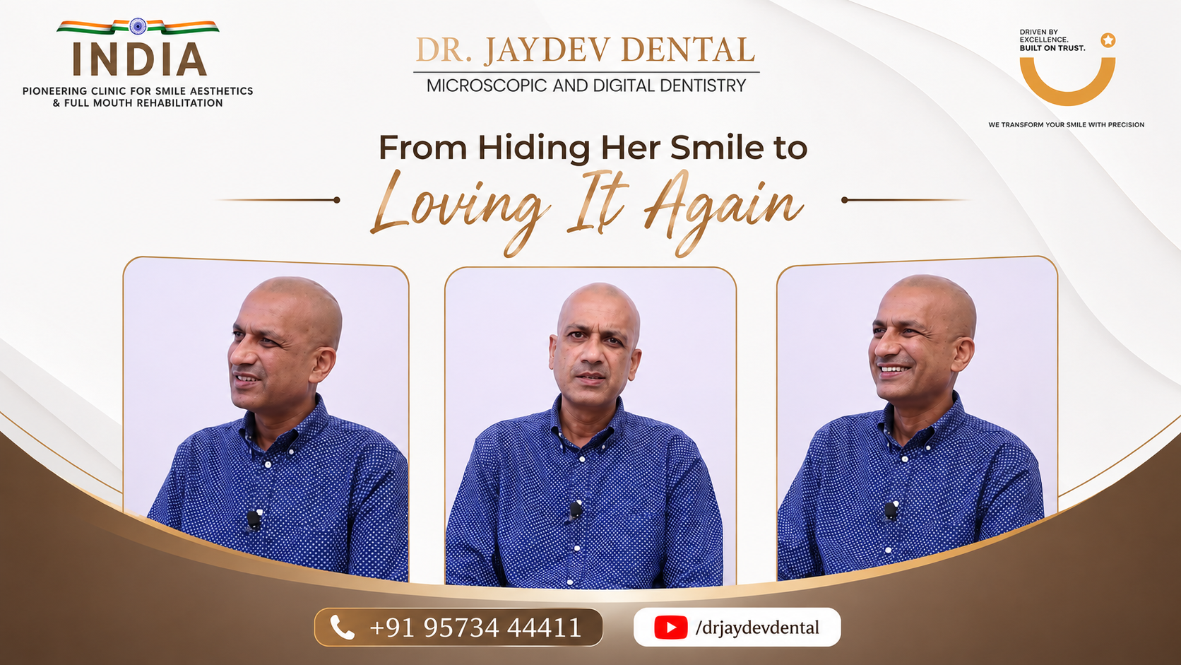
Watch the patient’s account in full:
The Patient's Concern
Three years before visiting Dr. Jaydev Dental Clinic, this patient developed severe dental pain and went to a local corporate hospital. The treating dentist took conventional X-rays and recommended root canal treatment on three teeth. The patient agreed and the procedures were carried out.
The pain never fully resolved. Even routine chewing caused discomfort that lasted for two and a half years. After living with the problem, the patient began searching specifically for a microscopic endodontist someone equipped to look deeper than a standard X-ray allows.
What the Patient Was Experiencing
→ Persistent pain while chewing across multiple teeth
→ Discomfort that had continued for approximately 2.5 to 3 years post-treatment
→ Three teeth with root canals that had not resolved symptoms
→ No clear explanation from the original treating clinic
The Challenge
The patient arrived having already undergone root canal treatment. This made the clinical situation significantly more complex than a first-time procedure. The primary goals were:
Determine precisely why all three treated teeth were still causing pain
Identify how much of the original canal system had been left untreated
Remove existing filling material without damaging the remaining tooth structure
Complete the root canal treatment in full under microscopic visualisation
Restore each tooth with a crown strong enough to function long-term
Diagnosis: What the 3D X-Ray Revealed
Dr. Jaydev began with a 3D CBCT scan (cone beam computed tomography), which provides a three-dimensional view of the entire root and surrounding bone that conventional X-rays cannot match. The findings were clear.
| Tooth | What the Previous Clinic Did | What the CBCT Scan Found |
| Tooth 1 | Root canal performed | 50% of pulp left inside the canal |
| Tooth 2 | Root canal performed | 50% of pulp left inside the canal |
| Tooth 3 | Root canal performed | 70% of pulp left inside the canal |
On every tooth, the pulp had not been fully removed. The canals were incompletely debrided, the source of infection remained, and the teeth were sealed over an untreated root system. The diagnosis was a complete failure of all three previous root canal procedures, a finding that explained every month of the patient’s pain.
Dr. Jaydev displayed the scan on a monitor and walked the patient through exactly what had happened and why. For the first time, the patient understood the cause of the problem and what it would take to correct it.
Why Microscopic Endodontics Was the Only Option
Retreatment of failed root canals is among the most technically demanding procedures in dentistry. The existing filling material must be removed without damaging the root, and the canal system, often curved, branched, or partly calcified, must be fully visualised and cleaned. This is not reliably possible without a dental operating microscope.
Dr. Jaydev Matapathi is one of India’s foremost microscopic endodontists and has trained dentists from across the country through hands-on workshops. His approach to re-root canal treatment combines 3D imaging for diagnosis with full microscopic visualisation during the procedure itself, a combination that reduces the risk of leaving anatomy untreated.
The Treatment Process
All three teeth were retreated in a structured sequence designed to eliminate infection and restore full function.
Step 1: 3D CBCT Imaging
A cone beam CT scan of all three teeth provided a full three-dimensional view of the root anatomy, existing filling material, and surrounding bone, giving Dr. Jaydev a precise map of what needed to be corrected on each tooth before beginning treatment.
Step 2: Removal of Existing Filling Material
Under dental operating microscope magnification, the failed filling material was carefully removed from each canal. The microscope allowed Dr. Jaydev to distinguish between filling material and tooth structure at every step, protecting the integrity of the roots.
Step 3: Complete Canal Debridement
Once the canals were cleared, the remaining pulp tissue, left untreated during the original procedures, was located, cleaned, and removed under full microscopic visualisation. Irrigation protocols ensured thorough disinfection of each canal system.
Step 4: Obturation and Sealing
Each canal was sealed completely with biocompatible material to prevent reinfection. The seal was verified radiographically before the patient left the clinic.
Step 5: 3M Lava Crown Restoration
All three retreated teeth were restored with 3M Lava crowns, high-strength zirconia ceramic restorations that provide superior aesthetics and long-term durability. Annual documented X-rays confirmed full healing across all treated teeth.
The Outcome
The results were complete and sustained.
Zero pain on chewing for the first time in three years
All three teeth fully treated and confirmed healed on annual X-rays
3M Lava crowns seated on all retreated teeth
Patient rated the outcome 10 out of 10
“No one should suffer like me. I had pain for two and a half to three years. Dr. Jaydev showed me on the monitor exactly what happened. After he redid all three root canals and placed the crowns, I was able to chew food properly with zero pain. On a scale of 1 to 10, I would give him a 10.”
Patient, Microscopic Re-Root Canal Treatment, Dr. Jaydev Dental Clinic
What Makes This Case Special
3D CBCT imaging used to confirm the exact extent of each canal failure before retreatment began
Full dental operating microscope visualisation throughout all three retreatment procedures
Pulp tissue retained by the original clinic (50%, 50%, and 70% per tooth) fully identified and removed
All three teeth restored with 3M Lava zirconia crowns for long-term strength
Annual follow-up X-rays documented complete healing on every treated tooth
